2018 American Society of Head and Neck Radiology
About This CME Teaching Activity
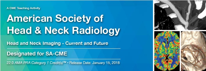
This CME teaching activity provides a broad review of head and neck imaging. Speakers present content-rich, clinically relevant materials on head and neck anatomy, nodal metastases, head and neck cancer staging, cranial nerve imaging, sinonasal imaging, and skull base imaging. In addition, pediatric head and neck imaging, parathyroid imaging, and surveillance imaging for head and neck cancer patients is included. Material contained in this product was captured during the 2014 ASHNR live meeting.
Target Audience
This CME teaching activity is designed to educate radiologists, neuroradiologists, dental and oral-maxillofacial practitioner, ENT physicians, head and neck surgeons, medical and radiation oncologists, general surgeons with other healthcare professionals interested in head and neck pathology, treatment and imaging.
Educational Objectives
At the completion of this CME teaching activity, you should be able to:
- Identify the anatomy of temporal bone, skull base, suprahyoid neck, and infrahyoid neck.
- Describe the lymphatic drainage and patterns of metastases.
- Review the anatomy of cranial nerves and clinical presentation of various pathologies affecting cranial nerves.
- Identify the imaging features and clinical significance of perineural tumor spread.
- Describe the common infectious and inflammatory process that involve the sinonasal region.
- Describe the clinical applications and indications of perfusion and diffusion imaging of head and neck tumor.
- Review current strategies for dose reduction for CT examination for head and neck.
- Discuss the benefit and limitations of PET/CT imaging for head and neck cancer.
- Recognize imaging features of various developmental and odontogenic maxillofacial lesions.
Details : 49 Videos
Size : 3.58 Gb
Price : $ 30
[WD_Button id=441]