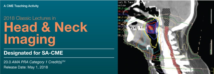
2018 Classic Lectures in Head and Neck Imaging
About This CME Teaching Activity
This CME activity brings together some of our most popular lectures in head & neck imaging. It combines a practical yet comprehensive review of head & neck imaging procedures concentrating on the latest trends, protocols and advances in clinical diagnosis, interpretation strategies and patient management. Faculty share techniques, tips and pitfalls through case based presentations.
Target Audience
This CME activity should be of interest to radiologists, otolaryngologists, and referring physicians who order these types of studies.
Educational Objectives
At the completion of this CME teaching activity, you should be able to:
- Identify the anatomy of temporal bone, skull base, suprahyoid neck, and infrahyoid neck.
- Review anatomy and common pathology of the cranial nerves and brachial plexus.
- Identify the imaging features and clinical significance of perineural tumor spread.
- Review the anatomy, features and pathology of the lymphatic system.
- Recognize image features of lymphadenopathy.
- Describe the patterns of tumor spread associated with nasopharyngeal cancers.
- Determine the optimal imaging techniques for evaluating head and neck pathology.
- Describe obstacles associated with post-treatment imaging in head and neck cancer, and how to minimize them.
Details : 35 Videos
Size : 4.13 Gb
Price : $ 30
Watch Samples[WD_Button id=441]