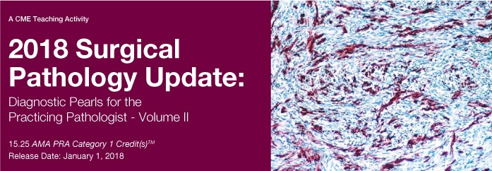
Surgical Pathology Update: Diagnostic Pearls for the Practicing Pathologist 2018
About This CME Teaching Activity
This CME Activity is designed to provide a comprehensive review of soft tissue, gastrointestinal, genitourinary, pancreaticobiliary and endocrine pathology. Content focuses on diagnostic pearls which lead to an accurate diagnosis as well as how to best avoid diagnostic pitfalls. In addition, faculty highlight the most useful and pertinent diagnostic immunohistochemical and molecular genetic techniques used in practice today.
Target Audience
This CME activity is primarily intended and designed to educate pathologists.
Educational Objectives
At the completion of this CME teaching activity, you should be able to:
- Develop an approach to common morphologic patterns seen in soft tissue tumors.
- Discuss the ancillary diagnostic approach to difficult soft tissue tumors including immunohistochemistry, molecular, and genetic diagnostic tests.
- Describe the most important diagnostic clues and patterns seen in soft tissue tumors which lead to a specific diagnosis.
- Explain the controversies regarding diagnostic criteria for Barrett’s esophagus and Barrett’s-related dysplasia.
- Discuss the effects of contemporary therapy on the pathology and management of patients with inflammatory bowel disease.
- Describe the role of the pathologist in helping to manage patients with IBD-related dysplasia and neoplasia.
- Apply risk stratification criteria for the prognostication of gastrointestinal stromal tumors.
- Discuss and recognize the succinate dehydrogenase-deficient gastrointestinal stromal tumor and understand the clinical significance of this category of GIST.
- Describe the transcription factors most useful for the identification of primary site location in metastatic carcinomas.
- Discuss the diagnostic markers for distinguishing hepatocellular carcinoma from metastatic carcinomas to the liver.
- Apply immunohistochemical markers to determine the primary site for metastatic well-differentiated neuroendocrine tumor.
- Accurately render a diagnosis of non-invasive follicular thyroid neoplasms with papillary-like nuclear features and recognize its mimics.
- Identify tumors with progression to poorly differentiated or anaplastic thyroid carcinoma.
- Recognize the morphologic spectrum of adrenal cortical carcinoma.
- Describe the features helpful in establishing a malignant diagnosis of prostate cancer on needle biopsy.
- Review the morphologic features essential in distinguishing benign mimickers from bladder cancer.
- Recognize unusual variants of common renal tumors and become familiar with more recently described renal tumors.
- Appreciate the subtle histomorphologic features that can help to distinguish well-differentiated ductal adenocarcinoma from reactive changes in the pancreas.
- Appreciate the anatomic and histologic compartments of the ampulla and their significance in the versatility and subclassification of ampullary tumors.
- Acquire knowledge of new concepts, terminology and histologic grading of pancreatic neuroendocrine tumors.
Details : 18 Videos
Size : 4.6 Gb
Price : $ 40
Watch Samples[WD_Button id=441]