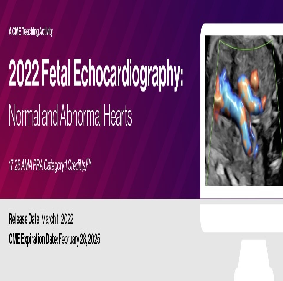
2022 Fetal Echocardiography: Normal and Abnormal Hearts

About This CME Teaching Activity
This CME activity provides detailed information on the evaluation of the normal fetal heart both from an anatomic and functional approach. In keeping with progress in this field, detailed information on the use of three-dimensional ultrasound in fetal echocardiography and cardiac imaging in the early gestation is presented. The CME activity also provides comprehensive evaluation of various cardiac malformations, involving abnormalities of the cardiac chambers and the outflow tracts.
Target Audience
This CME activity will greatly benefit physicians and sonographers in the fields of obstetric imaging, fetal echocardiography and pediatric cardiology. Residents in obstetrics and gynecology, as well as fellows in maternal fetal medicine and pediatric cardiology, will also benefit greatly. It should also prove valuable to those physicians and sonographers wanting to gain a more in-depth knowledge of fetal echocardiography.
Educational Objectives
At the completion of this CME teaching activity, you should be able to:
- Discuss indications for fetal echocardiography, review existing guidelines and the approach to patient counseling.
- Optimize ultrasound equipment for the use of 2D and Doppler in fetal echocardiography.
- Review the normal fetal cardiac anatomy.
- Describe the abnormalities of the venous system of the fetal heart.
- Discuss in detail anomalies of the cardiac chambers to include septal defects and anomalies of the right and left ventricle.
- Review in detail anomalies of the outflow tracts to include transposition of great arteries, tetralogy of Fallot complex and aortic arch abnormalities.
No special educational preparation is required for this CME activity.
Details : 39 Videos
Price : $ 75
[WD_Button id=441]